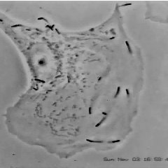
What do Listeria monocytogenes (a Gram positive rod), Shigella dysenteria (a Gram negative rod), and the Vaccinia virus (a double stranded DNA poxvirus) all have in common? They all hijack the actin polymerization capability of the host cell in order to become motile.
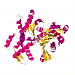
Actin is a globular, somewhat “U” shaped molecule that is used for many different functions in the cell (a moonlighting molecule). When actin exists as a monomer, it is known as G-actin, short for Globular actin. The aspect of actin that is exploited by the pathogens is the ability of actin to form long, branched, fibers.
When G-actin is polymerized into a fiber, it is known as F-actin, short for Filamentous actin. F-actin is polarized, with one end barbed and the other end pointed. The F-actin fiber grows from the barbed end and tends to dissociate at the pointed end. Growth of the fiber is accomplished by having an ATP bound G-actin bind to the barbed end. About two seconds after binding to the fiber, the ATP is hydrolyzed to ADP, and about six minutes after binding the ADP-G-actin complex dissociates from the F-actin filament. The rate of fiber extension or dissolution can be changed by cellular enzymes, thereby giving the cell control of the fiber growth.
Fibers can also branch by using Actin Related Proteins (ARP) 2 and 3. ARP2/3 binds to a fiber and starts the growth of a new fiber. This branching creates a tangle of fibers that has a large amount of drag in the intracellular environment. By extending F-actin from the tangle of fibers a force can be generated that can act upon an object. Using an enzyme that catalyzes the extension reaction, a force can be generated in one direction. In this way, the growing tangle of F-actin pushes the enzyme coated object forward. Mammalian cells use the enzyme profilin placed upon a cell membrane to create a pseudopod structure.
It is important to note that the F-actin fibers themselves do not move, but simply continue to elongate and thereby pushes the catalytic object. Because the F-actin dissociates after a while, the tangle spontaneously dissolves. The released G-actin molecules can be reactivated by ATP and can then move back to the head of the fiber.

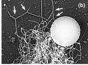
Polystyrene bead with ActA coating on 37.5% of surface area showing F-actin filament formation associated with ActA
Lysteria monocytogenes uses a different enzyme, Actin assembly-inducing protein (ActA), to induce the extension of F-actin filaments. By expressing ActA at only one end, the rod shaped L. monocytogenes is able to be pushed forward. As seen in the attached images, the pushing action can be replicated by coating a polystyrene bead with ActA on one side and putting the coated bead in a cell free solution that contains G-actin and ATP.
The pushing action induced by Listeria monocytogenes is shown in this excellent video from the lab of Dr. Julie Theriot by Julie Theriot & Dan Portnoy.
For a more complete review of this topic, I heartily suggest Dr. Julie Theriot’s lecture series at iBioSeminars, especially Part 1: Cell Motility. Her 44 minute lecture will give you a relatively complete understanding of how cells move – an understanding that is truly critical to understanding the world around us.
Here is a YouTube video of Vaccina virus moving inside a cell. The Vaccinia virus is labelled with green fluorescence and the actin filaments are red. Unlike L. monocytogenes, Vaccinia does not have a strongly polarized coating of F-actin polymerizing enzyme.
Another excellent reference is the work of the Liu Research Group, headed up by Dr. Andrea Liu.
Clearly, this has been a cursory summary of this important topic. My thanks go out to all of those researchers who have spent their precious time placing bricks on the tower of knowledge.


